The Shroud of Turin proves the Resurrection
Photography of the Shroud of Turin
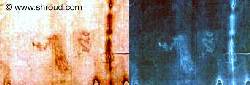
 The first photograph of the Shroud in 1898
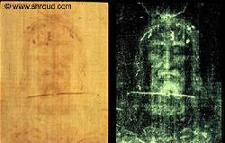
Interest in the Shroud dramatically increased in 1898 when the Italian photographer Secundo Pia took the first photograph of the Shroud in 1898. Both pictures above have a mixture of photo-positive and photo-negative images, which we will study in detail.
The picture on the left is a photo-positive picture of the face on the Shroud, with red blood marks from the Crown of Thorns. There is a faint image of a face, which is in photo-negative, making it difficult to understand.
To Secundo Pia’s astonishment the photo-negative face image on the Shroud (above left) became a photo-positive image on his camera negative (above right), except that the blood was in photo-negative, and therefore appears white on the black and white negative. The image of the Crucified Man in photo-positive is now much more clearly seen!
The photo-negative image (above left)
The image above left is a photo-positive image, with actual blood marks clearly seen in red, arising from the scalp wounds caused by the crown of thorns. The image of the face is in photo-negative, and is difficult to understand.
The photo-positive image (above right)
After Secondo Pia had photographed the photo-negative image of the face (above left) in 1898 he developed in his laboratory the amazing image of Jesus Christ on the right, in true photo-positive, with the blood marks more clearly seen in white, in photo-negative.
Both pictures have a mixture of photo-positive and photo-negative features, which we will study in detail.
The blood stains on the Shroud
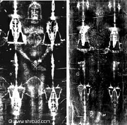
 The Shroud has blood marks on the scalp, both wrists, the right chest wall, and both ankles. This photograph of the Shroud clearly shows blood marks (in white) over the scalp, both wrists (with blood flows from both puncture wounds), blood flows from both plantar surfaces of the feet (see the dorsal image below right), and multiple laceration wounds from the Scourging.
The blood has been analysed. It is Type AB, the rarest blood group, and also contains a high level of Bilirubin. This is a bile pigment, which is found in high levels in the blood of torture victims. The bile pigment Bilirubin is bright red. This red colour is maintained post mortem, and accounts for the bright colouration of the blood on the Shroud.
The Scourging
 Multiple dumbbell shaped lacerations can be seen over the back, upper and lower aspects of both legs, and the anterior chest wall. According to Dr Robert Bucklin M.D. a Professor of Pathology, and former forensic pathologist, there are over 120 separate lashes, performed by two separate soldiers. Each single lash produced multiple lacerations, with severe injuries to the skin and underlying soft tissue. This was a most severe Scourging, and Jesus Christ was nearly scourged to death.
On the photograph of the dorsal image can be clearly seen the scourge marks over the back, and lower limbs. On both images can be clearly seen the profuse bleeding from the wounds in the scalp, and from both wounds in the feet, in white photo-negative. This is because the Blood of Jesus Christ actually stained the Shroud, before the Image was created at His Resurrection.
The Crown of Thorns
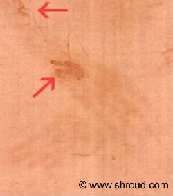
 On the photograph on the right, multiple rivulets of blood can be seen tracking from the scalp. This was almost certainly caused by the “crown of thorns”, which is one of the ways by which the victim on the Shroud is identified, since this was a torture reserved only for Jesus Christ. The crown of thorns was almost certainly a bush of thorns from the Lote bush, which grows wild in the Jerusalem area. The thorns are up to two inches long, and caused profuse bleeding.
The crown of thorns is recorded in Mark 15:17-18: “And they clothed Him with purple; and they twisted a crown of thorns, put it on His head, and began to salute Him, “Hail, King of the Jews!”
Abrasion to tip of nose
Forensic examination of the Shroud by Dr Robert Bucklin (see below), and other pathologists reveal the abrasion to the tip of the nose. There is also bruising of the right cheek, almost certainly caused by beating and punching.
The nailing of the hands
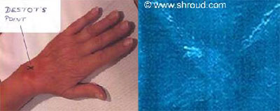 Examination of the area of the hands on the Shroud shows the left hand over the right hand (remember the image is transposed in a photo – negative image). There is also a puncture wound in both wrists, from which blood is seeping, draining up both forearms on the Shroud.
In mediaeval paintings the nails were always painted inserted into the palms, rather than the wrists. Experiments on dead bodies shows that the weight of a human body cannot be supported by nails through both palms.
As the Shroud clearly shows, the nails were in fact inserted through the wrist, through a small depression easily palpable on the dorsum of the wrist known as “Destot’s point”. The Roman soldiers were very skilled at Crucifixion, and searched for this exact point. The nails would have passed directly through the Median nerve in the Carpal Tunnel, causing excruciating pain.
As can be clearly seen on the Shroud, the thumbs appear to be missing from both hands. There is a reason for this, as explained below.
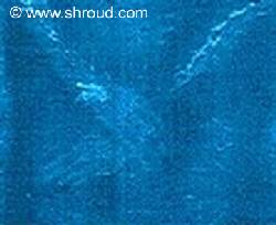
The position of the arms on the Cross
 We believe that the correct position of the arms on the Cross is as shown, with the palms against the wood of the Cross (the Patibulum) and the thumbs pointing downwards.
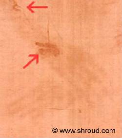
Traditionally the Crucifixion has always been displayed with the nails passing though the palms, and the dorsum of the hand against the crossbar. This is not correct. The blood flow from the wounds in the wrists shows that when the arms were placed on the Cross, they were placed with the palms against the wood of the Cross, with both thumbs pointing downwards.
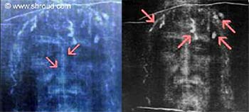
The blood trails can be clearly seen with the blood flowing on the dorsum (the back) of the forearms, from medial to lateral (on the back of the forearm, this is from the side of the forearm with the little finger, to the side of the forearm with the thumb. Try it on yourself!)
The blood flows from the injuries in the wrist indicate that the victim died with His hands raised about 65 degrees from the horizontal on the Cross. (To define medial and lateral, in the “anatomical position” the palms are facing forward).
In the blood flows shown in the picture to the right, the blood on the dorsum of both forearms is clearly tracking from medial to lateral. The only way this could have occurred is if the arms were positioned on the Cross with palms against the wood of the Cross, with both thumbs pointing down to the feet, and the nails hammered into Destot’s points.
The significance of this is that after six hours on the Cross, the weight of Jesus’ body would have dislocated His shoulders, elbows and wrists. Jesus died with His body supported by both arms, raised about 65 degrees from the horizontal. The arms are six inches longer than normal on the Shroud, due to the dislocations.
 In addition, Jesus’ body weight would have forcibly flexed both thumbs into the palms, and Jesus’ hands would have adopted a “claw” position. This is the reason why the thumbs are not seen on the Shroud. It is inconceivable that a mediaeval forger would be familiar with these anatomical details, which are certainly not well understood today, judging by all the anatomically incorrect images of the Crucifixion.
The wound between the 5th and 6th right ribs
There is a large blood stain over the right chest wall area, consistent with the post mortem wound to the chest. There is a penetrating skin wound in the right chest wall produced by a sharp puncturing instrument, almost certainly a Roman spear. The spear wound is between the 5th and 6th Right ribs.
Dr Robert Bucklin comments: “This wound has all the characteristics of a post-mortem type flow of blood from a body cavity or from an organ such as the heart. At the upper plane of the wound is an ovoid skin defect which is characteristic of a penetrating track produced by a sharp puncturing instrument.”
The nailing of the feet
 The image below left is the probable position in which the feet where nailed. The image below right is an X-ray showing the probable position of the nail, between the 2nd and 3rd Metatarsal spaces.
The image below left shows the blood red colouration on the Shroud where the feet were positioned. The image below right shows the same area photographed, with the blood as a photo-negative white colouration.
www.shroud.com